BRJ Medical
Peripheral catheter displacement can begin with a small movement. A sleeve pulls against the extension set. A patient bends the wrist. Tape lifts after handwashing. Within hours, the catheter may migrate, leak, or irritate the vein. In 2026, preventing this problem requires more than adding adhesive. It requires careful assessment, appropriate stabilization, and regular observation.
Vascular-access expert Nancy Moureau emphasizes, “Stabilize the catheter to protect the vein.” Her message reflects a practical clinical principle. Securement should limit catheter motion without restricting circulation or damaging fragile skin. How to secure a peripheral catheter to prevent displacement? Start by selecting a suitable site, cleansing the skin correctly, and allowing it to dry completely. Use a sterile, skin-compatible stabilization device when available. Keep the hub supported. Create a gentle loop in the extension tubing, rather than allowing tension to reach the insertion site.
Check the dressing edges.
Do not ignore small changes.
A loose border, damp adhesive, blood at the site, or new tenderness deserves attention. Nurses should reassess after insertion, patient movement, transfers, and bathing. Documentation also matters because it reveals repeated failures, such as wrist placement or poor tubing alignment. Still, no method works perfectly for every patient. Sweaty skin, edema, confusion, and frequent movement can defeat a well-applied dressing. That limitation deserves honesty. Effective prevention combines evidence-based technique with patient-specific judgment, timely replacement, and continuous reassessment.
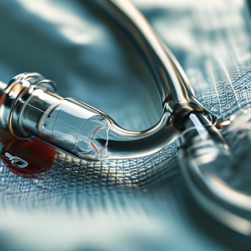
Peripheral catheter displacement means the device no longer sits securely inside the vein. Its tip may withdraw, migrate, or press against the vessel wall. Infusion can then slow, stop, or leak into surrounding tissue. The skin may look puffy, cool, pale, or tender. Sometimes, the first sign is a pump alarm.
International evidence shows this is not a minor bedside inconvenience. A large analysis of 11,830 catheters reported that about four in ten failed before treatment ended (Marsh et al., International Journal of Nursing Studies, 2018). Dislodgement was one recognized cause, alongside occlusion, phlebitis, and infiltration. The Infusion Nurses Society’s 2024 Standards of Practice also highlights securement, site assessment, and patient movement. Definitions still vary between studies.
Movement is often the quiet culprit. A restless arm, repeated elbow bending, or tubing pulled across a bedsheet can create tiny traction. Sweat, oily skin, fragile tissue, and loose dressings reduce stability. Children, older adults, and patients with edema need closer observation. In practice, rushed assessments can miss early lifting at the dressing edge. That is uncomfortable to admit. A catheter can appear usable while its tip has already shifted. Check for pain, swelling, resistance, dampness, and a changed infusion rate; never rely on appearance alone.
2026 How to Prevent Peripheral Catheter Displacement?
How to Assess Risks Before Peripheral Catheter Placement
Before insertion, assess more than vein visibility. Ask whether the patient is restless, confused, diaphoretic, oedematous, or likely to move the limb. Inspect skin integrity, circulation, sensation, and previous access sites. Small clues matter. Choose the smallest suitable catheter and a site away from joints when therapy allows. The Infusion Therapy Standards of Practice, 9th edition (2024), emphasizes matching catheter selection with therapy, vein condition, and patient factors.
A 2018 international study examined 40,638 peripheral catheters across 51 hospitals. It found that 36% failed before treatment ended, according to the Journal of Vascular Access. Dislodgement was one failure pathway. Infiltration, occlusion, and phlebitis often shared similar preventable conditions. Before placement, check whether the prescribed solution may irritate fragile veins. Consider the patient’s mobility, skin moisture, and ability to report discomfort. Not all risks show.
Risk assessment should continue after skin preparation. Check fixation needs, tubing direction, and whether movement could pull the catheter. CDC infection-control guidance supports regular site assessment and prompt removal when complications appear. Do not wait for swelling. Document vein depth, selected site, catheter size, patient cooperation, and reassessment time. Yet assessment is not perfect. A calm patient may suddenly flex an elbow during transfer. I have seen secure dressings fail when tubing pulled against a bedrail. Recheck after movement, transfer, or analgesia. Placement decisions improve when clinicians challenge their first impression.
How to Prevent Peripheral Catheter Displacement?
How to Insert and Secure a Peripheral Catheter Properly
Peripheral catheter stability begins before the dressing is applied. Perform hand hygiene, confirm the patient and site, and choose the smallest suitable gauge. Prepare the skin with an approved antiseptic and allow it to dry completely. Insert bevel-up until blood return appears. Lower the angle, advance the catheter, then release the tourniquet. Never reinsert a needle into the catheter. Small details matter.
After insertion, flush according to local policy and check for pain, swelling, leakage, or resistance. Use a sterile, transparent dressing and a stabilization method that limits movement at the hub. Keep the extension tubing looped gently, without pulling against the skin. Label the dressing with the date and time. The CDC recommends aseptic technique, routine site assessment, and removal when complications occur. The Infusion Nurses Society also emphasizes securement and regular evaluation.
An international observational study of more than 40,000 peripheral catheters across 51 countries reported failure in roughly one-third of devices before removal (Alexandrou et al., 2018). Dislodgement was one contributing problem. A perfect-looking dressing can still hide early migration. Nurses should inspect the insertion site at every appropriate contact, especially after transfers, bathing, or vigorous movement. In practice, rushed securement remains an avoidable weakness. Recheck technique through competency audits, because habits can drift quietly.
Clinical application: Select the smallest catheter gauge that meets the treatment requirement. Larger-bore catheters generally provide higher flow, but catheter selection should also consider vein size, therapy type, and patient condition.
To reduce displacement, perform hand hygiene, prepare the skin with an appropriate antiseptic, allow the site to dry completely, avoid touching the prepared area, advance the catheter gently after flashback, and secure it with a sterile stabilization dressing. Keep the hub supported and avoid placing tension on the extension tubing.
Data note: The flow rates shown are representative nominal values commonly published for peripheral catheters; actual performance varies with catheter design, needleless connectors, tubing, fluid viscosity, and pressure.
Peripheral catheter monitoring begins with a careful look at the insertion site. Check it before each use and during routine rounds. Compare the skin with the previous assessment. Early warning signs include new pain, tenderness, swelling, redness, coolness, or dampness beneath the dressing. The infusion may slow unexpectedly. The patient may report burning or pressure. Small changes matter.
Keep the dressing clean, dry, and fully visible. Do not cover the site with bulky materials that hide the catheter. Check whether the extension tubing pulls against the skin during movement. Ask the patient to report discomfort immediately. A loose dressing, visible catheter movement, or changed catheter length requires prompt clinical review.
Never force a flush against resistance. Stop the infusion and follow local clinical protocols.
I have sometimes underestimated mild tenderness because the skin looked normal. That was a mistake worth remembering. Appearance alone cannot confirm catheter stability. Reassess after repositioning the patient, transferring them, or changing the infusion.
Document the site condition, patient feedback, and any action taken. When findings are unclear, seek assessment from a trained clinician rather than guessing. Clear communication between shifts also prevents missed changes.
When peripheral catheter displacement occurs, stop the infusion immediately. Do not flush the line. A displaced catheter can push fluid into surrounding tissue, causing swelling, pain, cool skin, or blanching. Compare both limbs, check the dressing, and inspect the insertion site carefully. If the catheter has moved outward, never push it back in. Remove it according to local policy, apply gentle pressure, and insert a new catheter only when clinically necessary. Suspected vesicant extravasation requires urgent escalation and drug-specific guidance.
Act early. A large systematic review reported peripheral intravenous catheter failure in about 36% of adult catheters, with dislodgement among the preventable causes (Marsh et al., 2021). The Infusion Nurses Society’s 2024 Standards also emphasize ongoing site assessment, securement, and prompt action when complications appear. Record the time, symptoms, infusion solution, catheter condition, and care provided. Inform the responsible clinician, especially when swelling spreads or neurovascular changes develop. Watch for increasing redness, drainage, fever, numbness, or reduced movement.
My practical mistake was once trusting a clean dressing too quickly. It looked secure, but the catheter had shifted during patient movement. A visible dressing is not proof of stability. Reassess after transfers, bathing, and painful repositioning. Use gentle stabilization and avoid tension from tubing. Local protocols differ, and that matters. When uncertain, pause and ask an experienced clinician rather than guessing.
| Clinical Dimension | Common Risk or Warning Sign | Prevention Measure | What to Do When Displacement Occurs | Required Follow-up |
|---|---|---|---|---|
| Site selection | Catheter placed near a joint, in an area of frequent movement, or in a site with poor visibility. | Choose an intact vein with good blood flow; whenever clinically appropriate, prefer an upper-extremity site and avoid areas of flexion. | Stop using the catheter if the external length has changed, the dressing is loose, or the device appears partially withdrawn. | Assess the site and the affected limb for pain, swelling, redness, leakage, coolness, or changes in skin color. |
| Securement and dressing | Loose edges, wet or soiled dressing, visible catheter movement, or tension on the extension tubing. | Use sterile, transparent, semipermeable dressing and an appropriate needleless securement method; keep tubing supported without pulling on the catheter. | Do not push a partially withdrawn catheter back into the vein. Stabilize the limb and notify a qualified clinician. | Replace the catheter if its position or sterility is uncertain, according to local policy and clinical assessment. |
| Patient movement | Repeated bending of the elbow or wrist, pulling during transfers, restless movement, or accidental snagging. | Explain the purpose of the catheter, secure tubing during transfers, and position the limb comfortably while preserving circulation and mobility. | Pause the infusion if displacement is suspected and inspect the catheter, dressing, tubing, and insertion site. | Re-educate the patient and caregivers about avoiding traction, twisting, or manipulating the catheter. |
| Infusion assessment | Resistance to flushing, an infusion pump alarm, slowed flow, leakage, or an unexpected change in infusion pressure. | Check catheter patency and the insertion site before each use and at regular intervals during therapy, following institutional policy. | Do not force-flush. Stop the infusion, disconnect safely if required, and assess for infiltration, extravasation, occlusion, or phlebitis. | Follow the specific extravasation or infiltration protocol when a vesicant, irritant, or high-risk solution may be involved. |
| Signs of infiltration | Swelling, cool or pale skin, blanching, tightness, discomfort, or fluid leaking into surrounding tissue. | Inspect the site frequently and ask the patient about pain, burning, tightness, or unusual sensations during infusion. | Stop the infusion immediately. Do not flush. Aspirate residual medication only when directed by the applicable protocol, then remove or manage the catheter as clinically indicated. | Measure and document the affected area, elevate the limb when appropriate, and provide treatment based on the infused solution and local protocol. |
| Signs of phlebitis or infection | Pain or tenderness along the vein, erythema, warmth, palpable cord, drainage, fever, or spreading redness. | Use hand hygiene and aseptic technique, disinfect access points correctly, and replace dressings that are damp, loose, or visibly soiled. | Stop using the catheter and notify the responsible clinician. Remove the catheter when infection or phlebitis is suspected, unless a specific clinical protocol states otherwise. | Document findings and monitor for progression; obtain cultures or further evaluation only when clinically ordered. |
| Accidental complete removal | Catheter is no longer in the vein, bleeding is present, or the device is found detached from the dressing. | Use securement appropriate to the patient’s activity level and keep the insertion site visible for routine inspection. | Apply gentle pressure with sterile gauze until bleeding stops, cover the site, and assess the catheter to confirm it is intact. | Document the event, inspect for a retained catheter fragment, assess the patient, and insert a new catheter only if ongoing therapy is required. |
| High-risk medication or solution | Displacement occurs during infusion of a vesicant, irritant, hyperosmolar solution, or medication requiring reliable vascular access. | Confirm vascular access suitability before administration and use enhanced monitoring required by the medication protocol. | Stop administration immediately, leave the catheter in place initially if aspiration or antidote delivery may be required, and activate the extravasation protocol. | Record the medication, concentration, estimated amount, symptoms, interventions, and patient response; arrange specialist review when indicated. |
| Documentation and quality review | Repeated dislodgement, unclear insertion records, or delayed recognition of site complications. | Record insertion date and time, site, catheter gauge and length when required, number of attempts, securement, patient education, and ongoing assessments. | Escalate unexpected events through the facility’s clinical incident or vascular-access reporting process. | Review contributing factors such as site, securement, patient activity, therapy type, and assessment frequency to reduce recurrence. |
Safety note: Peripheral catheter assessment and treatment should be performed by trained healthcare professionals in accordance with current institutional policies and the medication-specific protocol.
: Perform hand hygiene and confirm the patient and insertion site. Choose the smallest suitable catheter gauge. Clean the skin with an approved antiseptic. Allow the skin to dry completely before insertion. Dry skin matters.
Insert the catheter bevel-up until blood return appears. Lower the angle and advance the catheter carefully. Release the tourniquet afterward. Never reinsert the needle into the catheter. Small details matter.
Flush it according to local policy. Use a sterile, transparent dressing. Add stabilization that limits movement near the hub. Loop the extension tubing gently. Avoid pulling against the skin.
Label the dressing with the insertion date and time. Keep the site visible for routine inspection. Avoid bulky coverings that hide the catheter. A clear view helps.
Watch for new pain, tenderness, swelling, redness, or cool skin. Dampness beneath the dressing may also signal a problem. The catheter may appear longer or visibly shifted. The infusion might slow unexpectedly. Small changes count.
Never force a flush against resistance. Stop the infusion and assess the site. Check for pain, swelling, leakage, and catheter movement. Follow local clinical protocols and seek trained assessment. Do not guess.
Check the site before each use and during routine rounds. Reassess after bathing, transfers, repositioning, or vigorous movement. Compare the current skin appearance with earlier findings. Ask the patient about burning, pressure, or discomfort.
No. A neat dressing can still hide early catheter migration. I have underestimated mild tenderness when the skin looked normal. That judgment was wrong. Appearance alone cannot confirm stability. Document findings and communicate changes between shifts.
Peripheral catheter displacement occurs when the catheter shifts, bends, backs out, or leaves the vein, potentially causing ineffective therapy, discomfort, infiltration, or tissue injury. This article explains how to identify risks before placement, including patient movement, fragile veins, insertion-site location, skin condition, and the planned duration of treatment. It also outlines careful vein selection, aseptic insertion, appropriate stabilization, and clear dressing techniques. A key question is: How to secure a peripheral catheter to prevent displacement? The answer involves using a suitable securement method, keeping the insertion site visible, avoiding excessive tension on the tubing, and allowing enough flexibility for normal movement without pulling on the catheter.
After placement, regular observation is essential. Healthcare professionals should check the site and catheter function for pain, swelling, redness, leakage, looseness, resistance, or changes in infusion flow. If displacement is suspected, stop the infusion, assess the patient and site, follow local clinical procedures, and replace or remove the catheter when necessary. Consistent documentation and patient education can further reduce complications.