 BRJ Medical
BRJ Medical
BRJ Medical
China Best IV Catheter Care How to Prevent Phlebitis? Safe catheter care begins with consistent clinical assessment, not a single product choice. Phlebitis can develop when the vein becomes irritated, inflamed, or infected. Early signs may include warmth, redness, tenderness, swelling, or a firm vein along the catheter path. The patient may simply say, “It feels tight.” That detail matters.
How to manage an IV catheter site to prevent phlebitis? Begin with hand hygiene and follow your facility’s approved aseptic technique. Inspect the site before each infusion and whenever the patient reports discomfort. Check the dressing for moisture, lifting edges, blood, or contamination. Keep the catheter stabilized, because repeated movement can irritate the vessel wall. Use the prescribed flushing method and compatible solutions. Never ignore resistance, leakage, or a sudden change in infusion flow.
Remove or replace the catheter according to clinical findings and local policy. Do not routinely continue using a painful or inflamed site. Escalate suspected phlebitis to a qualified healthcare professional promptly. Document the site appearance, symptoms, interventions, and patient response. Evidence-based practice supports regular observation, but real-world care is rarely perfect. Lighting may be poor. Documentation may be delayed. A busy ward can miss subtle redness. That is why simple reassessment matters. Clear communication between nurses, patients, and caregivers strengthens safety. When unsure, pause the infusion and seek clinical guidance rather than guessing.
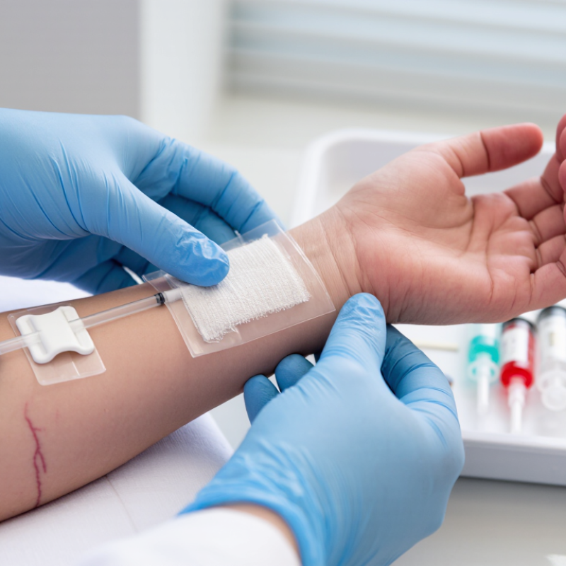
Phlebitis is inflammation of a vein after intravenous catheter placement. The area may become red, warm, swollen, or tender along the vein. Some patients describe a burning sensation when fluid enters. A firm, cord-like vein can appear in more advanced cases. Not every red mark is phlebitis. Tape irritation, bruising, infiltration, and infection can look similar. Careful assessment matters.
Phlebitis may develop from mechanical, chemical, or infectious causes. A catheter that moves inside the vein can scrape the vessel wall, especially near the wrist or a bending joint. Concentrated or irritating medicines may inflame the lining when diluted poorly or infused too quickly. Bacteria can enter through poor hand hygiene, contaminated equipment, or loose dressings. It can happen quietly. Early checks should include pain, temperature, color, swelling, and infusion flow. A practical bedside lesson is to compare both limbs, not only the insertion site. The vein may look normal while tenderness spreads upward. Catheter size, placement technique, vein condition, and dwell time also influence risk. Older adults, dehydrated patients, and people with fragile veins need closer observation. My assessment may still be imperfect; a busy shift can make subtle changes easy to miss. When phlebitis is suspected, stop using the catheter and follow the facility’s clinical protocol for removal, evaluation, and documentation. Persistent redness, drainage, fever, or worsening pain requires prompt medical review.
| Data Dimension | Definition or Indicator | Evidence-Based Information | Prevention or Care Action | Clinical Priority |
|---|---|---|---|---|
| Condition | Phlebitis | Inflammation of a vein that may occur near or along the path of a peripheral IV catheter. It can be mechanical, chemical, or infectious. | Inspect the catheter site and the surrounding vein regularly, and document any changes in appearance, pain, or function. | Requires prompt assessment |
| Early clinical signs | Localized irritation | Pain, tenderness, erythema, warmth, swelling, induration, or a palpable cord along the vein may indicate developing phlebitis. | Stop using the site if symptoms are present, assess the patient, and follow the facility's IV catheter removal and replacement protocol. | Act early |
| Mechanical cause | Physical irritation of the vessel wall | Movement of the catheter, excessive catheter size, poor stabilization, insertion near a joint, or repeated manipulation can traumatize the vein. | Choose the smallest suitable catheter, avoid areas of flexion when possible, use aseptic insertion technique, and stabilize the catheter securely. | Preventable risk |
| Chemical cause | Infusate-related irritation | Extremes of pH or osmolarity, high infusion concentration, and some medications can irritate the vascular endothelium. | Check medication compatibility and administration guidance, dilute or infuse according to clinical instructions, and monitor the site during administration. | Review infusion plan |
| Infectious cause | Microbial contamination | Infection may occur when microorganisms enter through the insertion site, contaminated equipment, poor hand hygiene, or inadequate skin antisepsis. | Perform hand hygiene, use appropriate skin antisepsis, maintain aseptic non-touch technique, keep the dressing clean and intact, and replace it when damp, loose, or soiled. | Urgent evaluation |
| Patient-related factors | Individual susceptibility | Small or fragile veins, dehydration, poor peripheral circulation, advanced age, a history of difficult access, and repeated cannulation can increase risk. | Assess the patient and veins before insertion, maintain hydration when clinically appropriate, limit unnecessary attempts, and escalate difficult access according to local policy. | Individualized care |
| Insertion site | Vein and location selection | Upper-extremity veins are generally preferred for adult peripheral IV access. Areas of flexion, infected skin, wounds, and sites distal to complications should be avoided. | Select a healthy vein, use the most distal appropriate site first, and avoid placing the catheter through areas of irritation or compromised skin. | Plan before insertion |
| Catheter selection | Size and material suitability | A catheter should be selected according to the prescribed therapy, flow requirement, vein condition, and expected duration. A smaller suitable catheter generally causes less vessel trauma. | Use the smallest gauge and shortest length that safely meet the clinical requirement, while following local standards and product instructions. | Optimize selection |
| Securement | Catheter stability | Catheter movement can create repeated friction against the vein wall and may increase mechanical irritation and accidental dislodgement. | Use a sterile, transparent, semipermeable dressing or an approved securement method; keep the insertion site visible whenever possible. | Maintain stability |
| Site monitoring | Routine assessment | The site should be assessed at regular intervals and whenever the patient reports pain, burning, tightness, leakage, or discomfort during infusion. | Compare the site with the opposite limb when helpful, check for blood return or patency only according to local policy, and record assessment findings. | Monitor continuously |
| Infusion technique | Administration conditions | Rapid infusion, excessive pressure, incompatible solutions, or incorrect flushing may contribute to discomfort, infiltration, or vessel irritation. | Use the prescribed rate, verify compatibility, flush with the approved solution and volume according to policy, and never force a resistant catheter. | Follow protocol |
| Response to suspected phlebitis | Immediate management | Continued infusion through a painful, red, swollen, or cord-like vein may worsen tissue and vascular injury. | Stop the infusion, assess the patient and catheter site, remove and replace the catheter when indicated, notify the responsible clinician, and document the event. | Immediate action |
| Escalation signs | Possible infection or serious complication | Fever, purulent drainage, rapidly spreading redness, severe pain, red streaking, marked swelling, or systemic symptoms require urgent clinical assessment. | Stop using the catheter and obtain immediate medical evaluation according to the healthcare facility's escalation procedure. | Urgent clinical review |
| Quality indicator | Phlebitis rate | Phlebitis rate is commonly expressed as the number of phlebitis cases divided by the number of peripheral IV catheters or catheter-days, multiplied by 100 or 1,000. | Use a consistent case definition, collect data prospectively, review trends, and combine surveillance results with staff education and process improvement. | Track trends |
Key practice point: Phlebitis is not always preventable, but early vein assessment, appropriate catheter selection, strict aseptic technique, secure stabilization, suitable infusion practices, and regular site monitoring can reduce risk and support timely intervention.
Clinical note: Exact catheter replacement intervals, flushing procedures, skin antiseptics, and escalation criteria should follow the current policies of the healthcare facility and applicable national clinical guidelines.
Choose the smallest catheter that safely supports the prescribed therapy. A narrower catheter may reduce vein trauma. The choice is not always obvious.
Use good lighting to assess the veins. Prefer distal veins before proximal sites. Avoid bruised, swollen, infected, or irritated skin.
Warm the limb briefly. Do not repeatedly probe after a failed attempt. Repeated attempts can increase tissue injury.
Perform hand hygiene and use aseptic technique. Clean the skin with an approved antiseptic. Let the area dry fully. Do not touch it afterward.
Keep the bevel facing upward. Advance gently after seeing blood return. Never force the catheter against resistance. Resistance needs assessment.
Use a sterile dressing that keeps the insertion site visible. Avoid tight tape. Make a gentle tubing loop to reduce pulling during movement.
Use only the prescribed solution and volume. Follow local clinical policy and aseptic technique. Never force the flush. Resistance may signal blockage or displacement.
Check for pain, burning, warmth, redness, swelling, leakage, or a hardened vein. Stop the infusion if symptoms appear. Follow the approved clinical procedure.
Observe it before, during, and after infusion. Record pain, skin condition, dressing status, and catheter function. Routine checks can become careless, so reassessment matters.
Phlebitis is inflammation of a vein that may develop after IV catheter placement because of mechanical irritation, chemical irritation, poor site selection, or inadequate hygiene. How to manage an IV catheter site to prevent phlebitis? Begin by choosing the smallest suitable catheter and an appropriate, healthy vein, then insert it using aseptic technique with minimal manipulation. Secure the catheter without excessive pressure, keep the dressing clean, dry, and intact, and flush the line only according to approved clinical procedures.
Regular monitoring is essential. Check for redness, warmth, swelling, pain, tenderness, firmness, leaking, or changes in infusion flow. At the first sign of phlebitis or another complication, stop using the catheter, assess the patient, and follow facility protocols for removal, replacement, documentation, and medical evaluation. Prompt recognition, gentle handling, and consistent catheter care can reduce discomfort, prevent worsening inflammation, and support safer IV therapy.
